Posterior Lumbar Interbody Fusion (PLIF)
A posterior lumbar interbody fusion (PLIF) is a surgical technique that involves correction of the spinal problems at the base of the spine by placing bone graft between two vertebrae. Minimally invasive surgical techniques may be used to perform the procedure.
What is the Minimally Invasive PLIF surgical approach?
Posterior lumbar interbody fusion (PLIF) is a spinal surgery that involves placement of bone graft material between two adjacent vertebrae from the back of the spine. The bone graft material works as a bridge or platform that binds or fuses two vertebrae together and promotes new bone growth. The ultimate purpose of the procedure is to reestablish the spinal stability.
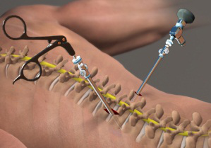
Now-a-days minimally invasive spine surgical techniques may be used to perform PLIF which permits the surgeon to make small incisions and gently separate the surrounding muscles of the spine rather than cutting. The aim of the minimally invasive approach is to preserves the surrounding muscular and vascular function for faster recovery and reduced scarring.
What are the indications for Minimally Invasive PLIF surgery?
 Patients with spinal instability in their lower back due to degenerative disc disease, spondylolisthesis or spinal stenosis that has not responded to other non-surgical treatment measures such as rest, physical therapy or medications may be recommended for surgical treatment of spinal fusion procedure such as PLIF. Patients with lumbar spinal instability may experience pain, numbness and muscle weakness in the lower back, hips and legs.
Before prescribing the PLIF surgery your surgeon considers various factors such as the condition to be treated, your age, health, lifestyle and your expected level of activity after the surgery. Have a complete discussion with your spinal care provider regarding the available treatment options.
What is the procedure for Minimally Invasive PLIF surgery?
In this procedure the surgeon makes a small incision in the lower back over the vertebra (e) to be treated. The size of the incision depends on the bone graft to be used and can be as small as approximately 3 centimeters. Usually 3- to 6- inches of incision is required in a traditional open PLIF surgical procedure.
The surgeon dilates the surrounding muscles of the spine to access the section of the spine to be stabilized. Next, the lamina, the roof of the vertebra, is removed to visualize the nerve roots. The facet joints that are directly over the nerve roots are trimmed to provide the nerve roots with more space.
Bone Graft Placement
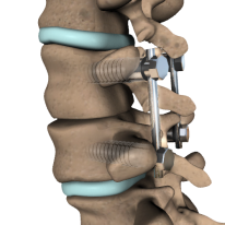
 To place the bone graft the nerve roots are shifted to one side and disc material is removed from the front (anterior) of the spine. Now the bone graft is implemented into the disc space. Screws and rods are used to stabilize the spine for better healing and fusion.
After the completion of the procedure the small incision is closed and leaving behind a minimal scar.
What is the recovery period following PLIF surgery?
This minimally invasive procedure typically permits most of the patients to be discharged the day after surgery but some patients may require a longer hospitalization. Usually most of the patients observe immediate improvement of some or all of their symptoms but sometimes the improvement of the symptoms may be gradual.
Contribution of a positive approach, realistic expectations and compliance with your doctor’s post-surgical instructions help bring a satisfactory outcome of the surgical procedure. Most patients can resume their regular activities within several weeks.
Have a conversation with the doctor to determine if you are a candidate for minimally invasive PLIF surgery.
What are the risks and complications of PLIF surgery?
Every patient has a particular treatment plan and outcome result. Sometimes outcome results vary from individual to individual. The complications associated with PLIF surgery include infection, nerve damage, blood clots or blood loss, bowel and bladder problems and problems associated with anesthesia. The primary risk of spinal fusion surgery is failure of fusion of vertebral bone and bone graft which may require an additional surgery.
Please refer to your physician to obtain an ample list of indications, warnings or adverse effects or precautions, clinical results and other significant medical information related to the minimally invasive PLIF surgical procedure.
Deformity Program


 Read More
 Location Map
 Patient Testimonials
 Insurances


